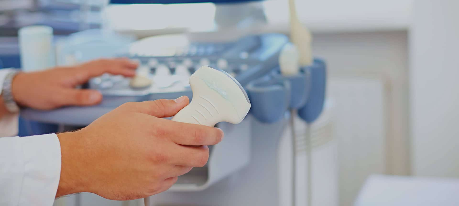
Carotid arterial assessment using non-invasive real time ultrasound scanning, combined with Doppler colour and spectral analysis is fast becoming the test of choice in determining haemodynamic occlusive disease. In fact, with the aid of experienced Technologists, many Vascular surgeons are now performing surgery based on duplex findings without the need for angiography and therefore avoiding its complications.
Carotid duplex scanning is a cost effective and very accurate screening test and gives the surgeon valuable information on the degree of stenosis and the type of plaque present . The vertebral arterial circulation may also be assessed before or after exercise in order to demonstrate subclavian steal.
Indications:
- Cervical bruit
- TIAs
- CVA
- Amaurosis fugax and scotoma
- Follow up after carotid endarterectomy or stenting
- Pre-operative evaluation
- Non-lateralising, less specific symptoms, may include dizziness, headaches and vertigo.
- Suspicion of pulsatile mass in carotid or subclavian region.
- Suspected subclavian steal syndrome (BP < 30mmHg in contralateral arm)
